Femur Bone
Largest bone in the body
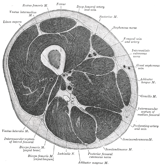
The Archive/Images/Gray anatomy 1918 images/Myology/image244 Right femur. Anterior surface.gif The Archive/Images/Gray anatomy 1918 images/Myology/image245 Right femur. Posterior surface.gif The Archive/Images/Gray anatomy 1918 images/Myology/image246 Lower extremity of right femur viewed from below.gif The Archive/Images/Gray anatomy 1918 images/Myology/image247 Frontal longitudinal midsection of upper femur.gif The Archive/Images/Gray anatomy 1918 images/Myology/image248 Diagram of the lines of stress in the upper femur, based upon the mathematical analysis of the right femur. These result from the combination of the different kinds of stresses at each point in the femur.gif The Archive/Images/Gray anatomy 1918 images/Myology/image249 Frontal longitudinal midsection of left femur. Taken from the same subject as the one that was analyzed and shown in Figs. 248 and 250. 4:9 of natural size.gif The Archive/Images/Gray anatomy 1918 images/Myology/image250 Diagram of the computed lines of maximum stress in the normal femur.gif The Archive/Images/Gray anatomy 1918 images/Myology/image251 Intensity of the maximum tensile and compressive stresses in the upper femur. Computed for the load of 100 pounds on the right femur.gif The Archive/Images/Gray anatomy 1918 images/Myology/image252 Plan of ossification of the femur. From five centers.gif The Archive/Images/Gray anatomy 1918 images/Myology/image253 Epiphysial lines of femur in a young adult. Anterior aspect. The lines of attachment of the articular capsules are in blue.gif The Archive/Images/Gray anatomy 1918 images/Myology/image254 Epiphysial lines of femur in a young adult. Posterior aspect. The lines of attachment of the articular capsules are in blue.gif
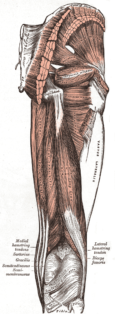
Muscles
Histology
Compact bone
The cortex of the femur contains mostly compact bone2. The compact bone tissue is “dense” and “unyielding,” and thus can withstand large loads especially shear and torsional forces2.
Cancellous bone
Cancellous bone is a relatively porous and spongy bone tissue that is made up of 3D trabecular lattices which span the inside of the femur2. Cancellous bone gives the femur its elastic properties, allowing it to repeatedly absorb external forces2.
Due to Wolff’s Law, cancellous bone will concentrate along lines of stress, resulting in defined trabecular networks2. The trabeculae is separated into the medial trabecular network and arcuate trabecular network2.
Osteologic features
Femoral head
The femoral head can be found just inferior to the middle 1/3 of the inguinal ligament2.
The femoral head is covered by articular cartilage except for the fova2.
Femoral Neck
The length of the femoral neck impacts the efficiency of the gluteus medius and glute min muscles3.
If there was no femoral neck, the available ROM of the hip would increase significantly, but the moment arm of the gluteus medius and gluteus minimus would shorter and thus its torque generation would be significantly less3.
Load-Absorption
The femur can withstand the high repetitive forces due to walking and other activities via its composition of compact bone and cancellous bone2.
Angle of Inclination (AoI)
The angle of inclination refers to the angle between the femoral neck and medial femoral shaft in the frontal plane2.
| Stage of Development | Angle of Inclination |
|---|---|
| Birth | 165-170 °2 |
| 2-8 y/o | Decreases by 2 °/yr2 |
| Adulthood | 125 °2 |
| Name | Angle |
|---|---|
| Coxa vara | <125 °2 |
| Normal | 125 °2 |
| Coxa Valga | >125 °2 |
Femoral Torsion
Femoral torsion refers to the relative twist between the femur’s shaft and neck in the transverse plane2.
| Name | Angle |
|---|---|
| Retroversion | <8 ° |
| Normal | 8-20 ° anteversion |
| Excessive Anteversion | >20 ° |
Femoral torsion can be measured via the Craig’s Test
Femoral condyles
These two condyles are not identical.
The medial condyle is narrower and projects more anteriorly than the lateral condyle.
Landmarks
Linea Aspera
- Origin:
Intertrochanteric line
- Origin:
Intertrochanteric Crest
Quadrate Tubercle
The quadrate tubercle refers to the distal attachment of the Quadratus Femoris muscle
Lesser Trochanter
The lesser trochanter projects posterior-medially from the inferior end of the intertrochanteric crest2. The lesser trochanter serves as the insertion for the Iliopsoas muscle, and is thus indirectly related to hip flexion and vertical L/S stabilization2.
Fracture
The majority of femoral fractures occur at the femoral neck in elderly individuals with predisposing conditions such as osteoporosis4.
Femoral shaft fracture can occur, but are less common and require extreme amounts of force, such as a motor vehicle accident4.
Stress Fracture
Femoral Neck Stress Fracture
Femoral Neck Stress Fracture: ?var:ref-femoral-neck-stress-fracture.definition
Caution
- Immediate weight bearing restriction to prevent further fracture progression or compromised bony stability
- 6-8 weeks and up to 14weeks for complex patients with poorer prognosis
Prognosis
Uncomplicated
Complicated
Compression Sided
Tension sided
Poor Prognosis
- Tension-sided
- age?
- >50% width of femoral nec
Weeks 0-8
- NWB
- OKC exercises to strengthen
Aquatic exercises can be used to achieve a therapeutic exercise volume while not jeopardizing healing osseous tissue
RTS
Uncomplicated compression side FNSFs and pubic rami stress fractures that are managed nonoperatively can typically return to running and athletic activity around 12 weeks after diagnosis
However, return-to-sport timeframes of up to 28 weeks have been reported for complicated, and slow-healing cases
Rehab
- Gradual PT supervised return-to-running5
- Glute Medius strengthening
- Frontal Plane pelvic stabilization
- Pelvic stability during loading
Tension-sided FNFS
Tension-sided patients have higher risk for displacement with weight bearing5. As a result, surgery with a percutaneous screw fixation is the recommended course of treatment
Surgical Management
- Tension-sided FNFS
- Patients with compression sided FNSFs that are greater than 50% of the width of the femoral neck are also considered candidates
- If a fracture becomes displaced, surgery should be performed to mitigate the potential for adverse effects including postoperative AVN and fixation failure
Rehab
Week 0-6
- NWB5
Week 7-12
- PWB5
3-6mo
- Return-to-sport can be achieved around 3-6 months after surgery but, can take up to a year in complicated cases with delayed healing5


